Brain Metabolism
18F-2-Fluoro-2-Deoxy-Glucose or FDG is radioactive molecule most commonly chosen in Positron Emission Tomography (PET) to study the metabolic state of cells in the body. The radioactive 18F isotope is generated by proton bombardment of 18O-enriched water which will knock out a neutron. The 18F isotope is then conjugated to deoxyglucose through a series of chemical reactions. Cells require glucose to maintain their metabolic state. Cells that are more active require more glucose than those that are less active. FDG is taken into these cells from the general circulatory supply in the same way as Glucose. Furthermore, FDG becomes sequestered or trapped within those cells that take it in. This leads to a build up of FDG within metabolically active cells which can be visualized in PET imaging as the 18F isotope decays. Clinically this is used for several applications but is most commonly used to image the distribution of cancer cells within the body, relying on the principle that these cells are highly metabolically active. In preclinical research it is commonly used to image the metabolic response in the brain to a variety of stimuli.
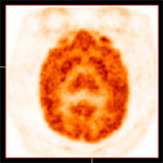
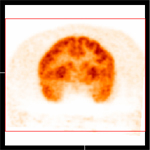
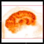
Transaxial View | Coronal View | Sagital View |
 |  |  |
The images above show the distribution of FDG uptake in chimpanzee (Pan troglodytes) brain. Distributions are shown in three different anatomic views.